About Us
The purpose of the club is to provide opportunity for members to enjoy golf in a social and supportive atmosphere.
Regular games are played over 9 holes on Monday, Wednesday and Friday mornings.
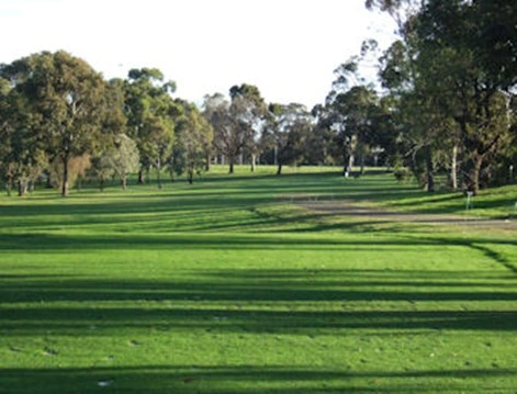
You will find us at the Royal Park Public Golf Course, located in Old Poplar Road, Parkville 3052
Membership
Application for membership can be made at any time using the RPVGC Membership Application
Membership fees for the club, payable from 1st September are:
{i} A one-off joining fee of $50 from 1st January 2024.
{ii} An Annual Membership fee of $95, payable from 1st September each year.
The Club is affiliated with Golf Australia.
Green fees for the golf course are applicable.
9 holes: Seniors $17.50
Annual green fees tickets are available from Melbourne City Council through the Pro Shop only during the month of July.
Included in the membership fees of the club are members benefits which include:
Free lunches
Free BBQ's
For all members there are also benefits with regard to:
Subsidised carts for those who need such assistance
Contact Us
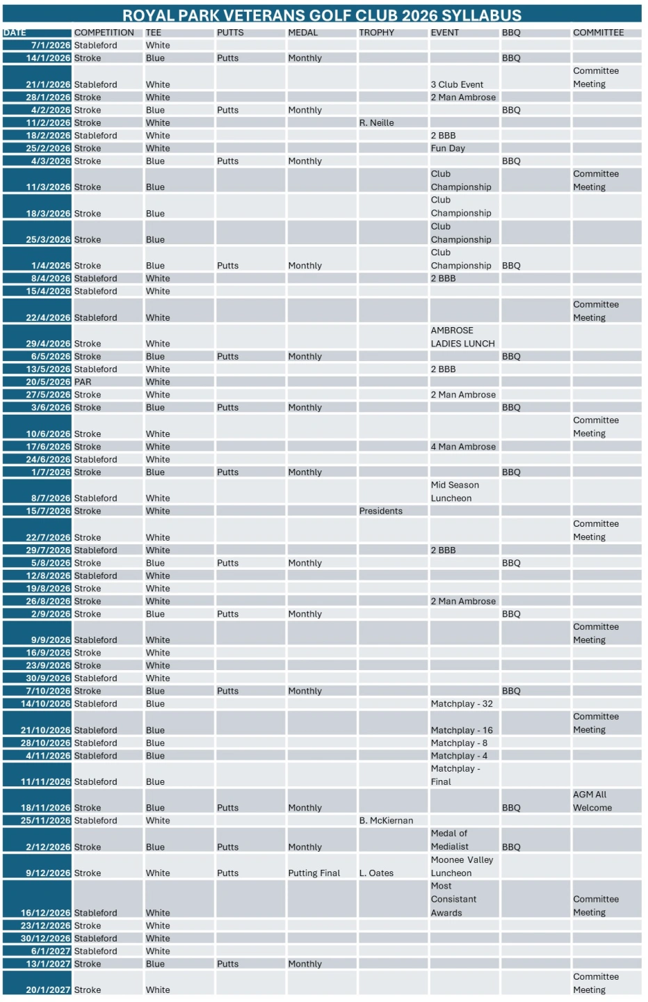
Syllabus RPVG 2026